Mitral Insufficiency

![]()
Mitral Insufficiency
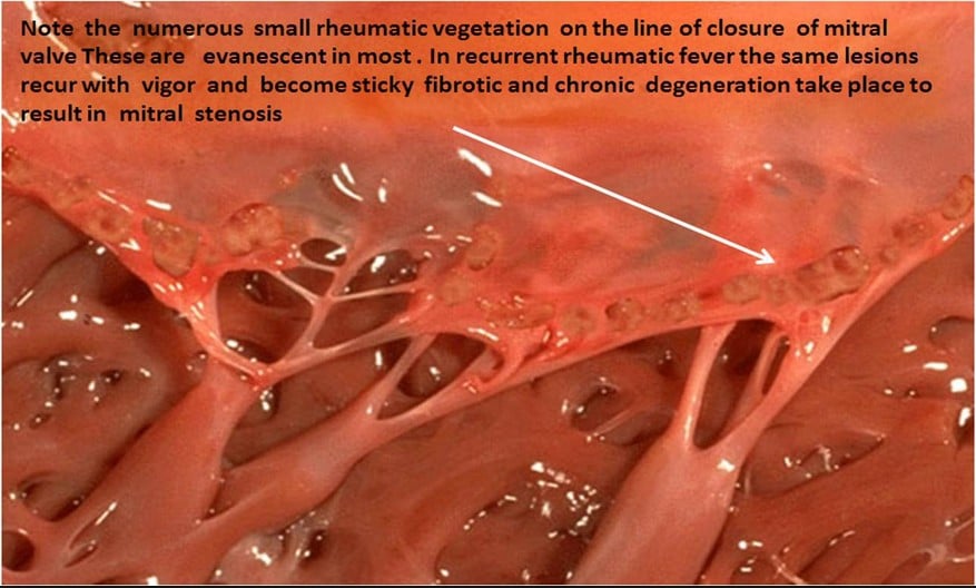
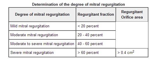
Six anatomical components must mesh synchronously to insure proper function of the mitral valve. Disruption of the kinetic interrelationship between the left ventricular wall, the left atrial wall, valve leaflets, annulus, chordae tendineae, and papillary muscles, may lead to dysfunction of the valve. Chronic mitral valve insufficiency (regurgitation) most often results from rheumatic fever. Other etiologies include mitral leaflet prolapse, coronary artery disease, left ventricular dilation, calcified mitral annulus, papillary muscle dysfunction related to infarction, and lupus erythematosus. The degree of regurgitation is established during cardiac catheterization and is defined by radiographic opacity of post-contraction residual dye initially injected into the left ventricle. This stratification design utilizes a 4-plus system, with 1+ representing mild regurgitation, and 4+ indicating the most severe state.

Assessment of left ventricular function based on ejection fraction may be misleading because of the low resistance exit state of the left ventricle. Essentially there are two compartments (directions) that become available for left ventricular volume to eject towards. Failing to identify and quantify volume that is directed back to the left atrium, may result in clinical misinterpretation of left ventricular function. While the fraction of volume ejecting from the ventricle is a traditional guideline for assessing ventricular function, this standard may be misleading in cases where a non-compliant ventricle may appear to empty normally. With mitral insufficiency, encountering patients with ejection fractions of less than 40% may indicate advanced myocardial dysfunction.
The severity of left atrial and ventricular dilation is directly proportional to the regurgitant stroke volume of the left ventricle. In acute cases, regurgitation into a normal non-compliant left atrium results in high pressures that may lead to pulmonary edema. Anxiety associated with dyspnea may potentiate this condition further by stimulating the sympathetic nervous system and aggravating the insult to the lungs by increasing systemic blood pressure.

Chronic mitral insufficiency allows for compensatory enlargement and compliance of the left atrium making it capable of handling ventricular backwash while preventing excessive pulmonary venous congestion. Right heart failure, atrial fibrillation and reduced cardiac output, will present if the condition remains untreated. The atrial filling phase is represented by the V wave in the pulmonary artery wedge tracing. These V waves become quite pronounced in cases of acute mitral insufficiency and represent retrograde ventricular ejection into the left atrium. A reduction of this manifestation can be realized using sodium nitropresside or nitroglycerin to promote vasodilation. Nitroglycerin has been demonstrated to reduce papillary muscle ischemia and anginal symptoms, while sodium nitropresside may improve forward cardiac output.
Treatment goals should be aimed to avoid circumstances that will increase regurgitation (bradycardia and hypertension), to optimize cardiac output (inotropic support), and to maintain hemodynamic equilibrium (keep the heart full).
![]()
References
Mastropietro C. Anesthesia for cardiac and peripheral vascular surgery. In Waugaman WR, Foster SD, Rigor, BM eds. Principles and Practice of Nurse Anesthesia. Norwalk, Appleton & Lange; 1992:705-748.
Ross AF, Gomez MN, Tinker JH. Anesthesia for adult cardiac procedures. In: Rogers MC, Tinker JH, Covino BG, Longnecker DE, eds. Principles and Practice of Anesthesiology. St. Louis, MO. Mosby Year Book; 1993;2:1649-1679.
Lewis KP. Early intervention of inotropic support in facilitating weaning from cardiopulmonary bypass: The New England Deaconess Hospital experience. Journal of Cardiothoracic and Vascular Anesthesia. 1993;7(Suppl 2):40-45.
![]()
